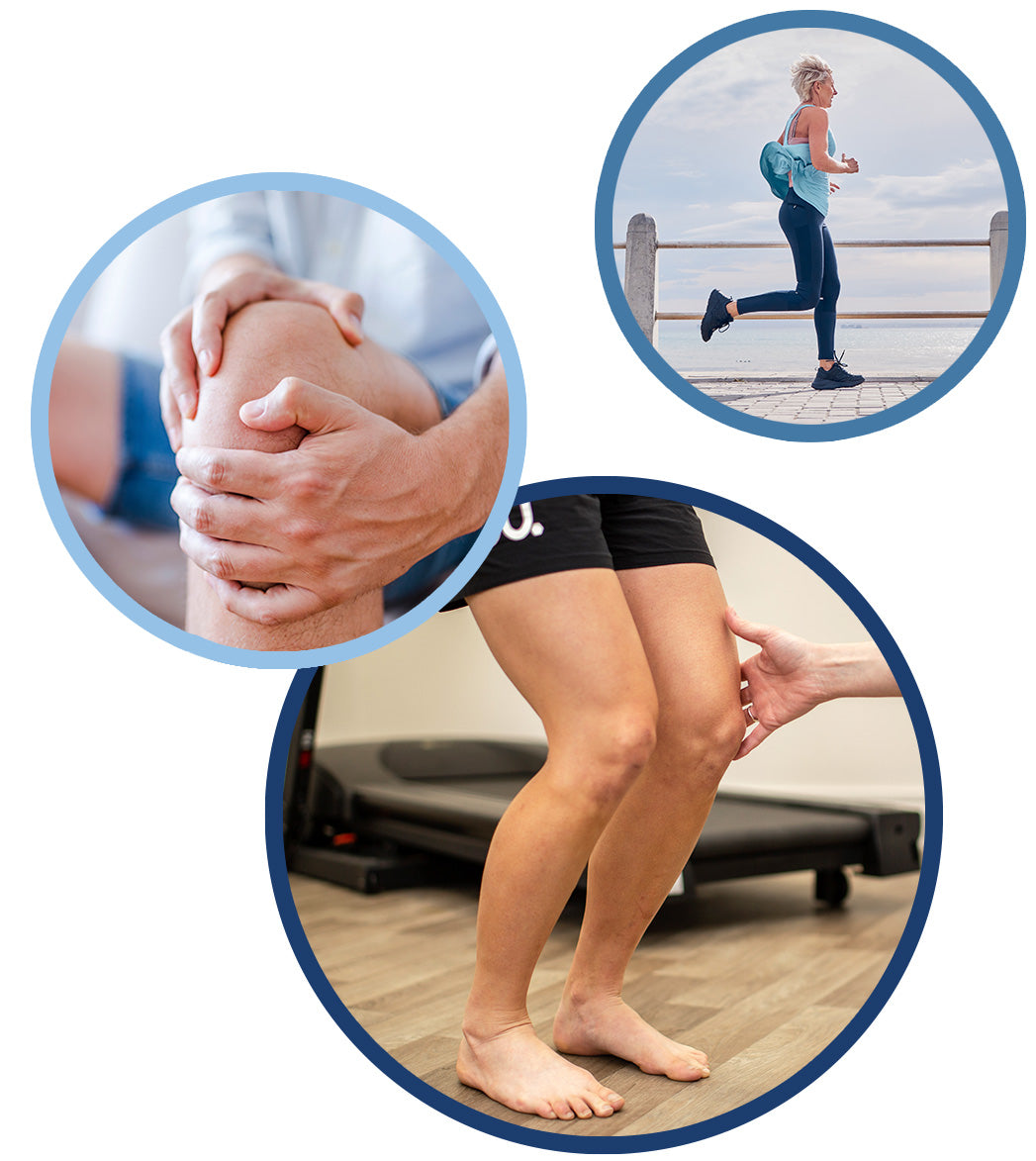
What's Causing My Knee Pain?
Knee pain: it’s a common problem for us Kiwis, and according to Australian research, the rate of knee injuries is on the rise - especially in kids aged 5-14 years,1 perhaps being attributed to growing participation rates in sports like soccer, basketball and netball that have an increased risk of knee injuries.
The incidence of knee pain can’t be a surprise - after all, our knee is the largest and most complex weight-bearing joint in our body. It takes on forces equivalent to 1.5 times your body weight when you’re walking across level ground,2 this rises exponentially during running to produce compressive forces of almost seven times our body weight on the tendon at the front of our knee, and up to eleven times our body weight on the patellofemoral joint, where the kneecap meets the thigh bone.
So what are the most common causes of knee pain that our podiatrists here at Merivale Podiatry see and treat? Here are seven to get started.
But First: An Overview
Before we dive into the causes, we thought we’d quickly give you an overview of the knee joint, as understanding the causes of knee pain is made much easier when you know more about how the knee joint works and all the parts within it. So: your knee is primarily made of three bones: the thigh bone (femur), the shin bone (tibia) and the kneecap (patella), which are enclosed in a joint capsule. The knee is a type of synovial joint, meaning that where the end of the femur and tibia meet inside the joint capsule, their surfaces are covered with a thin layer of strong smooth articular cartilage that works to help the bones glide seamlessly over one another as the joint flexes and straightens, while also helping to absorb shock. Over the articular cartilage is a thin layer of slippery joint fluid called the synovial fluid, which lubricates the cartilage-covered bone surfaces. Specifically, a healthy knee joint has up to 4ml of synovial fluid,3 which is just less than one teaspoon.
The kneecap sits outside of the joint capsule, protecting the front of the knee. Alongside the joint capsule, the knee joint is held in position and stabilised by an extensive network of ligaments, tendons and muscles.
Patellar Tendonitis
Patellar tendonitis describes inflammation and pain to the patellar tendon, the tendon that runs across the front of your knee, in which your kneecap is embedded. It is also nicknamed “jumper’s knee” because of the stress that is placed on the tendon during jumping sports, which is one of the primary causes of this condition. Your lower limb biomechanics, training technique and muscular imbalances such as tight hamstrings or weak quadriceps can also play a role in developing patellar tendonitis. We often see jumper’s knee in active adults involved in sports like netball or volleyball. You may notice:
- Knee pain and swelling around the front of the knee, particularly below the kneecap
- Pain that may develop gradually
- Stiffness and difficulty moving the knee, especially during activities that extend the knees like squatting
Patellofemoral Pain Syndrome
Patellofemoral pain syndrome (PFPS) describes pain at the point where the kneecap meets the thigh bone. It is colloquially known as “runner’s knee” because of its high prevalence in runners and those participating in running sports. It’s estimated to affect up to 23% of adults annually, affecting almost twice as many women as men.4 The painful symptoms at the knee develop when the kneecap mistracks, so that instead of gliding smoothly in a specific groove at the femur, it moves irregularly and rubs against the end of the femur as the knee bends and straightens. You may notice:
- Pain behind your kneecap and around your knee
- Swelling around the knee
- Symptoms may come on gradually during activities that repetitively bend and straighten the knee or put significant force through the knee, such as climbing stairs, squatting and running, as well as sports like basketball, skiing and netball
Anterior Cruciate Ligament (ACL) & Posterior Cruciate Ligament (PCL) Injuries
The ACL is one of two cruciate ligaments that sit within your knee joint and connect to both your shin bone (tibia) towards the front of the bone, and your thigh bone (femur) towards the back of the bone. The other ligament is the posterior cruciate ligament, and it also connects to both bones, but in the opposite direction, starting at the front of the femur and connecting to the back of the tibia. In this way, the two cruciate ligaments create a cross within the joint - hence the name “cruciate” ligaments. Both ligaments help to stabilise the knee and prevent excess movement from the femur on the tibia and vice versa.
We frequently see ACL injuries and tears in clients engaged in contact sports such as football, rugby, basketball, and skiing. An ACL injury is often caused by suddenly changing direction, twisting the legs and knees while the feet are firmly planted on the ground, or direct impact to the knee from a blow or during a collision, falling or landing poorly from a jump. You may notice:
- Sharp, sudden pain in the knee
- A sensation of popping, or a popping sound in the knee
- Swelling or stiffness in the knee
- Instability in the knee
- Difficulty walking
ACL injuries can also occur with damage to other ligaments and tendons in the knee, which is why it’s important to carry out a comprehensive assessment (often with ultrasound or MRI imaging) to understand the true extent of the injury and factor this into the treatment plan.
Posterior Cruciate Ligament (PCL) injuries are of a similar nature to ACL injuries, often occurring from a direct blow to the front of the knee during sporting activities or in an accident, including car accidents. The symptoms are similar to ACL injuries - feelings of instability or like your knee is giving out, pain and tenderness at the joint, swelling, and difficulty walking.

Collateral Ligament Injuries
While your cruciate ligaments (above) are found within the knee joint, the collateral ligaments are found at the sides of the knee. Specifically, you have the medial collateral ligament (MCL) on the inside of the knee that connects the femur to the tibia, and the lateral collateral ligament (LCL) on the outside of the knee that connects the femur to the fibula (the smaller bone in the lower leg).
These ligaments help to stabilise the knee and protect it from damaging sideways motion. Any tears or sprains to either collateral ligaments are usually a result of a direct blow to the outside of the knee, or due to a twisting force.
Torn Meniscus (Cartilage Damage)
Your menisci (singular = meniscus) are C-shaped rubbery cartilage discs present in pairs between the bones within the knee joint. They are known as the medial meniscus and the lateral meniscus depending on whether we’re referring to the meniscus on the inside of the knee (medial) or the outside of the knee (lateral). Menisci play an important role, working to absorb shock and distribute load through the joint, improve knee stability, and help control knee flexion and extension movements5 - to name a few.
We often see meniscal tears in young adults, often occurring as a result of high forces on the knees during sports - especially fast-paced sports that involve quick pivots or changes in direction, as well as those that require fast movements during activities that bend and straighten the knee. In older adults, it occurs due to the effects of ageing, including the thinning of the cartilage. When you injure a meniscus, you may notice:
- A popping sound
- Pain and swelling which progressively worsens over the 2-3 days following the initial injury
- Stiffness at the joint, or a feeling of the joint locking
- Instability of like the joint is giving way
- Bending the knee will exacerbate the symptoms
If your tear is degenerative in nature, then your symptoms may start with a dull ache or niggling at the knee before worsening.
Knee Bursitis
There are over 150 bursae in the body, including many around the knee. A bursa (singular) is a small, fluid-filled sac that sits between skin and tendon, or tendon and bone, to allow the tendons to glide smoothly and easily without rubbing. Bursitis occurs when any of the bursae in the knee become inflamed. It most frequently occurs on the inner side of the knee below the joint and over the knee cap. Bursitis can be a result of physical activity, trauma, infection, overuse, degeneration such as osteoarthritis, and more.6
Symptoms of knee bursitis can vary depending on which bursa is affected, but are usually described as sensations of pain, swelling and of localised heat. Symptoms also tend to start gradually and worsen over time - one of the many reasons why we encourage not ignoring knee aches and niggles, even if they’re mild.
Degenerative Changes – Osteoarthritis
Another common cause of knee pain we see is from degenerative changes to the knee joint. Natural age-related changes mean that our cartilage thins and the knee joint may begin to deteriorate. This means that the knee doesn’t move as smoothly as it used to, so the knee can feel painful and stiff. Osteoarthritic changes tend to develop over many years. You may feel:
- Stiffness in your knee
- Tenderness and swelling
- A creaking sound as the joint is moved
- Pain after sitting or resting, or that worsens with movement
If you’re living with knee pain, whether it’s persistent or tends to come and go without ever getting that “it’s gone for good” feeling, we highly recommend booking an appointment with one of our experienced podiatrists.
–––––––––––––––––––––––-–––––––––––––––––––––––––––––––––––––––
1 https://www.ncbi.nlm.nih.gov/pmc/articles/PMC8956823/
2 https://www.health.harvard.edu/pain/why-weight-matters-when-it-comes-to-joint-pain
3 Mundt L, Shanahan K MS MT(ASCP), Graf’s Textbook of Routine Urinalysis and Body Fluids (p.255), Philadelphia, Pa: Lippincott Williams & Wilkins; 2011
4 https://journals.plos.org/plosone/article?id=10.1371/journal.pone.0190892
5 Solomon L, Warwick D, Nayagam S. Apley's Concise System of Orthopaedics and Fractures. 3rd edn. Great Britain: Hodder Arnold, 2005.
6 https://www.ncbi.nlm.nih.gov/pmc/articles/PMC3354353
